
Electroencephalography (EEG) and magnetic resonance imaging (MRI) never yield identical results for the same patient. However, doctors combine these two techniques to obtain a more comprehensive view of brain activity.
The EEG measures electrical activity in real-time, while the MRI captures detailed images of internal structures. Each method has distinct advantages and specific limitations that guide their use in diagnosing or monitoring neurological conditions.
Further reading : Why Invest in Responsible Funds? Discover the Benefits You Can't Miss
EEG and MRI: two complementary approaches to explore brain activity
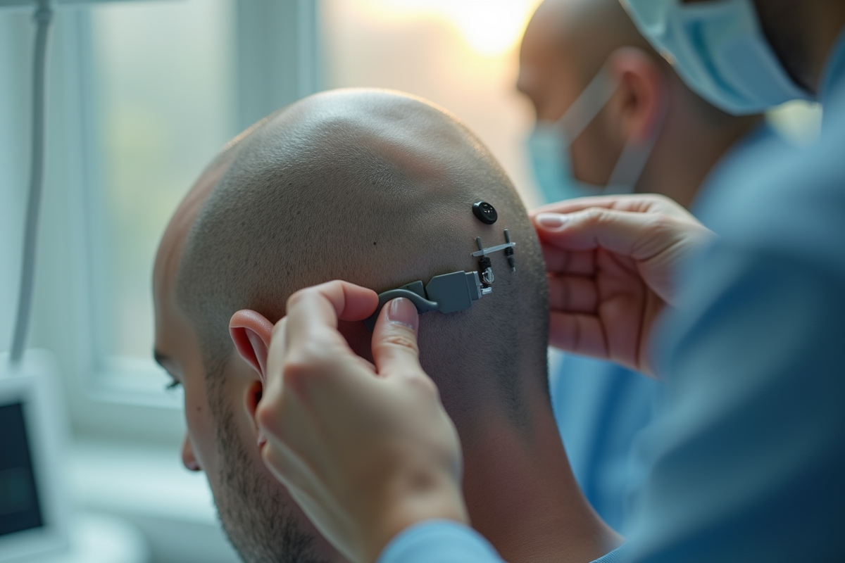
Exploring the brain means accepting that each tool reveals a different facet of its functioning. With electroencephalography (EEG), doctors delve into the brain’s electrical dynamics. Electrodes placed on the scalp continuously capture the dance of brain waves. Irregularities, discrete signals, or new rhythms may emerge, sometimes well before any symptoms manifest. The EEG provides real-time access to activity: it allows for the detection of vigilance disorders, the identification of the onset of a seizure, or the monitoring of sleep with great sensitivity.
In contrast, magnetic resonance imaging (MRI) offers an architect’s perspective. It maps the brain in minute detail: deep structures, brainstem, white matter, nothing escapes the scanner’s eye. The MRI detects lesions, identifies micro-hemorrhages, and reveals perfusion anomalies that would otherwise go undetected. Some early signals, such as Fazekas 1, serve as alerts. This discreet yet significant marker indicates the presence of white matter lesions even when the person feels nothing.
Related reading : Contemporary Artists Redefining the Codes of Modernity
Each of these methods has its strengths. The EEG captures the ephemeral, while the MRI immortalizes the traces left by time or disease. This complementarity goes beyond simple diagnosis: it guides monitoring in patients suffering from neurodegenerative diseases, informs therapeutic choices after a head injury or stroke. Observing the brain is learning to read movement and memory, to anticipate the unexpected, to document what, without these tools, would remain hidden.
What are the contributions, applications, and limitations of these examinations in medical practice?
Early detection of brain dysfunction can sometimes radically change a patient’s course. EEG and MRI are among the few examinations capable of revealing, from the earliest stages, anomalies that are completely undetectable during a simple consultation. Whether monitoring variations in consciousness, tracking sleep, analyzing reactions during a coma, or measuring the effects of a stroke, these techniques provide a unique view of the brain, an organ that still defies many certainties.
Here’s how these examinations are concretely integrated into medical monitoring:
- Measuring intracranial pressure or heart rate in fragile patients.
- Monitoring the progression of Alzheimer’s disease or Parkinson’s disease patients.
- Evaluating the consequences of a head injury on brain structure and function.
- Analyzing cerebrospinal fluid to complete the clinical picture.
However, the finesse of a trace or the clarity of an image is not everything. Interpreting an evoked potential, distinguishing a functional anomaly from a mere artifact, requires discernment. Limitations exist: the EEG, for example, does not always detect deep lesions, while the MRI is not accessible to everyone or at all times, especially in emergency situations or for unstable patients.
Technical expertise never supplants clinical insight. These examinations, powerful and valuable, do not replace the attentive listening of the patient or the physician’s holistic view. They enrich the understanding of the brain, this organ that continues to perplex and fascinate, but can never reveal everything on its own.
Monitoring early signals from the brain means accepting to walk a tightrope between medical intuition and cutting-edge technology. Sometimes, a tiny anomaly on a trace or an image changes a patient’s destiny. Other times, the real signal, the one that matters, resonates elsewhere: in speech, doubt, or the perseverance of a team. The brain still holds many secrets, but each examination, each exchanged glance, brings us a little closer.